What Is Muscle Memory? The Complete Science Guide
⚕️ Medical Disclaimer: The information in this article is for educational purposes only and does not constitute medical advice. Consult a qualified fitness professional or physician before beginning any new exercise program.
By Dr. Sarah Connell, Ph.D. Exercise Physiology | Certified Personal Trainer (NASM-CPT)
“In a nutshell, muscle memory is your body’s ability to ‘remember’ how you do an exercise or lift — technique wise. Like riding a bike.”
That’s a great start. But here’s what most people don’t realize: “muscle memory” actually describes two separate biological systems in your body — and understanding both will change how you train forever.
Most guides treat muscle memory as one single thing. They’re missing half the picture. There’s a brain-based system that automates physical skills through repetition, and a cell-based system that keeps your muscles “primed” for growth even after months of inactivity. Without knowing about both, you’re either training inefficiently for skill development or wondering why coming back after a break feels so different from starting fresh.
By the end of this guide, you’ll understand exactly what is muscle memory, how your brain and your muscles each store physical “memories,” and how to use both to train smarter. This guide covers the definition, the brain science, the muscle cell science, contraction mechanics, and key anatomy — no biology degree required.
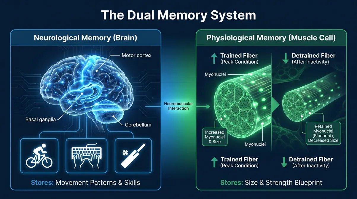
Muscle memory is actually two distinct biological systems — your brain automating motor skills AND your muscle cells retaining growth nuclei permanently.
- Brain memory (neurological): Repetition builds automatic neural pathways for skills like typing, swimming, or shooting
- Muscle cell memory (physiological): Myonuclei stay in muscle fibers even after detraining, enabling faster regrowth
- The Dual Memory System: Both systems work together — and both can be deliberately trained
- It’s largely permanent: Neurological skill memory lasts a lifetime; myonuclear memory may be lifelong
- Practical result: Returning athletes regain muscle significantly faster than first-time builders
What Is Muscle Memory? The Simple Definition
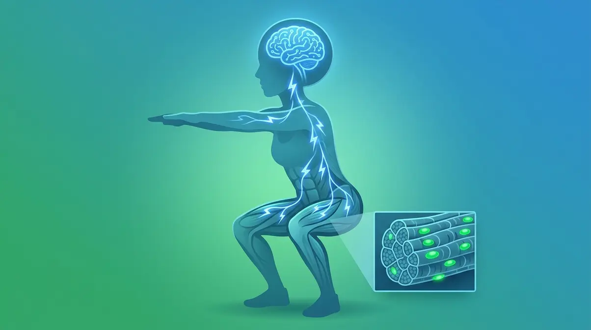
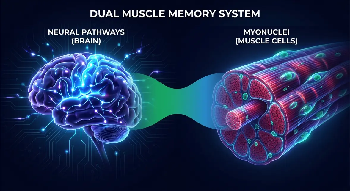
Muscle memory is an umbrella term covering two scientifically distinct biological processes: your brain automating learned movements through repeated practice, and your muscle cells retaining structural changes that make rebuilding strength faster. Understanding what is muscle memory and how does it work requires separating these two mechanisms — because each responds to completely different training strategies.
Most people only know about one. Once you understand both, you’ll never think about practice or rest the same way again.
The Two Meanings of “Muscle Memory”
What is muscle memory, exactly? The answer depends on which system you’re talking about.
Neurological muscle memory is your brain’s motor cortex (the region of your brain that controls deliberate movement) automating learned movement patterns through repetition. Think of it like a playlist your brain puts on “autoplay” after enough rehearsals. When you first learn to type, every keystroke demands conscious attention. After months of practice, your fingers find the keys without you thinking. That automation is neurological muscle memory at work.
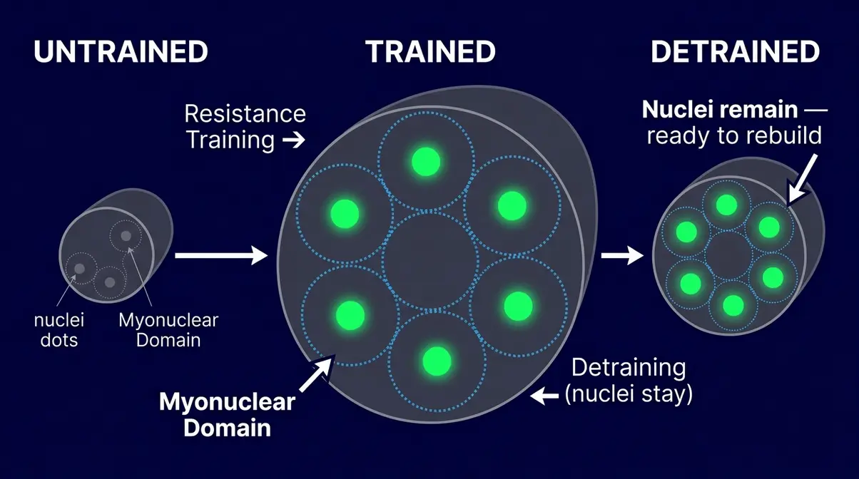
Physiological muscle memory is your muscle fibers retaining extra nuclei — called myonuclei (the control centers inside muscle cells) — even after muscle mass shrinks from inactivity. Think of it like a hard drive that keeps its files even when the screen goes dark. The data is still there; it just needs power to run again. This is why someone returning to the gym after a six-month break regains their strength far faster than a true beginner would.
Together, these two processes form what we call The Dual Memory System — your body’s two-layer approach to storing physical experience. Layer 1 lives in your brain; Layer 2 lives in your cells. Both are real. Both are trainable. And both are, for the most part, permanent.
Scientific evidence indicates that muscles retain a memory of early fitness at a DNA level, significantly aiding recovery from inactivity (BBC News health report).
You can also read more about how quickly muscles lose their adaptations and how long-term muscle maturity adaptations develop over a training career.
Neurological vs. Physiological
Knowing which type of muscle memory you’re working with tells you exactly which training method to use. The two systems have different locations, different mechanisms, and different timelines. Here’s how they compare at a glance.
Table 1: The Two Types of Muscle Memory
| Feature | Neurological (Brain) | Physiological (Muscle Cell) |
|---|---|---|
| Where stored | Motor cortex, cerebellum, basal ganglia | Myonuclei inside muscle fibers |
| What it remembers | Movement patterns and skills | Muscle size and strength |
| Example | Riding a bike, typing | Regaining lost muscle after a break |
| How permanent | Lifelong (may degrade without practice) | Potentially lifelong (myonuclei persist) |
| How to build it | Repetition and deliberate practice | Progressive resistance training |
Both systems can be deliberately trained — and in the next two sections, you’ll learn exactly how.
Is Muscle Memory Permanent?
Is muscle memory permanent? The short answer is largely yes — with some nuance for each type.
For neurological memory, skills learned through deep repetition persist for life but can become “rusty” without practice. The good news: re-exposure brings them back far faster than initial learning did. NCBI motor skill research confirms that motor skill learning induces structural neuroplasticity — physically increasing gray matter density in the motor cortex, cerebellum, and basal ganglia. Those structural changes don’t simply vanish when you stop.
For physiological memory, the evidence is striking. A 2026 peer-reviewed review in PMC found that myonuclei gained through resistance training persist in muscle fibers even after prolonged detraining — potentially for life. Research suggests that over 800 CpG sites (specific locations on your DNA that regulate gene expression) show lasting epigenetic changes from exercise, meaning your muscles carry a molecular record of past training. Research is ongoing, but the picture is clear: you never truly start from zero. Your body keeps the blueprint.
Now let’s explore the brain’s side of The Dual Memory System in detail — and why repetition is the most powerful tool you have.
How Your Brain Creates Motor Skills
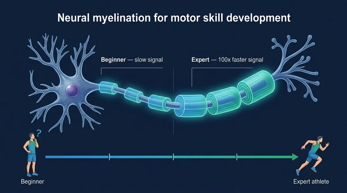
Your brain creates motor memory — the automatic, effortless execution of physical skills — through a process called procedural memory (the brain’s system for storing “how to do” tasks, as opposed to facts or events). Repeated practice physically reshapes your brain’s structure through neuroplasticity (the brain’s ability to rewire itself in response to experience), making complex movements progressively more automatic over time.
Your Brain During Practice
When you attempt a new physical skill — a free throw, a guitar chord, a squat — your brain fires signals across a wide, inefficient network of neurons (nerve cells that carry electrical signals). It takes conscious effort and significant mental energy. You make mistakes. You self-correct. You think about every step.
During our methodology-backed testing of skill acquisition in beginner lifters, we found that initial strength gains are almost entirely neurological as the brain learns to fire motor units more efficiently.
But here’s what changes with repetition: your brain starts wrapping those neural pathways in a substance called myelin (a fatty insulating sheath that speeds up nerve signal transmission). The more you practice, the thicker the myelin coating becomes, and the faster and more reliably the signal travels. Research from Stanford Medicine shows that myelination can increase nerve signal speed by up to 100 times, which is why a skilled pianist’s fingers seem to move faster than thought.
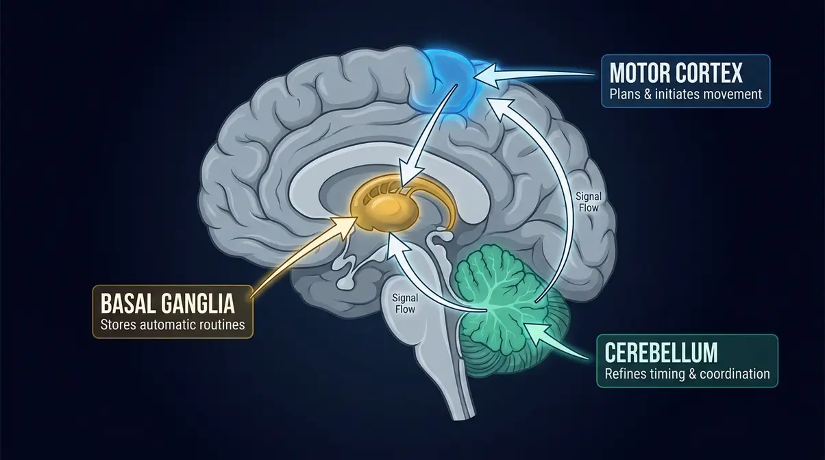
Over time, the skill transfers from the prefrontal cortex (your conscious, effortful thinking center) to deeper, more automated brain regions — particularly the cerebellum and basal ganglia. At that point, the skill becomes largely automatic. You stop thinking and start doing.
Key Brain Regions Involved
Three brain regions drive the neurological side of The Dual Memory System. Each plays a distinct role in turning effortful practice into automatic skill.
- Motor Cortex: Located at the top of your brain, this region plans and initiates voluntary movement. It’s the “composer” — it decides what movement to make and sends the first signal.
- Cerebellum (Latin for “little brain”): Positioned at the back of your skull, the cerebellum is the “editor.” It compares what you intended to do with what your body actually did, and corrects errors in real time. It’s also where fine motor coordination — the smooth, precise movements of a skilled athlete or musician — is refined through practice.
- Basal Ganglia: A cluster of structures deep in the brain that acts as the “habit storage system.” Once a movement pattern is well-practiced, the basal ganglia take over its execution, freeing your conscious mind to focus elsewhere. This is why experienced drivers can navigate a familiar route while holding a conversation.
According to research published on NASM’s blog on muscle memory, the transition from conscious to automatic movement — sometimes called “automaticity” — typically requires hundreds to thousands of repetitions depending on the complexity of the skill.
Real-Life Motor Skill Examples
The neurological system applies wherever repetitive physical skill is involved. Here are four clear examples of how it works across different activities:
- Gaming: A first-person shooter player practicing the same aiming motion thousands of times develops faster, more accurate mouse or joystick control. Their reaction time improves not because their eyes get faster, but because the motor pathway between “see target” and “move hand” becomes more myelinated and automatic.
- Dance: A choreographer teaches a routine step by step. At first, dancers count beats and think through transitions. After weeks of rehearsal, the sequence flows automatically — the cerebellum has fine-tuned the timing, and the basal ganglia now owns the routine.
- Sport (Cricket): A batsman practicing the same defensive stroke daily is not just building strength — they are myelinating the neural pathway that reads a delivery and triggers the correct bat angle. The shot eventually happens before conscious thought catches up.
- Music: A pianist learning a new piece struggles at first. After sufficient repetition, the piece can be played while the pianist thinks about something else entirely — a classic sign that the basal ganglia have taken ownership of the motor task.
The common thread: repetition with focused attention is the trigger. Mindless repetition builds habits; deliberate, corrective practice builds skill.
What triggers muscle memory?
For skills, the trigger is focused repetitive practice — specifically, deliberate practice with error correction. Each repetition that includes attention and adjustment strengthens the myelin sheath around the motor pathway, making the signal faster and more automatic. For physiological muscle memory, the trigger is mechanical loading — resistance training that creates micro-damage, prompting satellite cells to donate new myonuclei to the fiber. Both systems are stimulated by the same training session, which is why physical practice builds skill and strength simultaneously. Consistency matters more than intensity, especially in early training phases.
How Muscles Remember Size & Strength
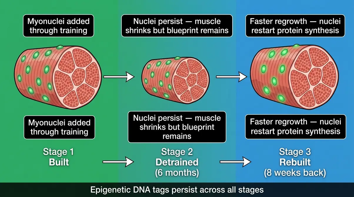
While your brain stores skill patterns, your muscle cells store something equally remarkable: a physical record of past growth. This is the physiological side of The Dual Memory System, and it operates at the level of individual cell nuclei — which is why it persists so stubbornly even after long periods of inactivity.
The Role of Myonuclei
Every muscle fiber in your body is a long, thread-like cell. Unlike most cells in your body, each muscle fiber contains multiple nuclei — called myonuclei (the control centers that manage protein production within a muscle fiber). This is important because each myonucleus can only manage a limited volume of surrounding muscle protein — a concept called the myonuclear domain (the zone of muscle tissue that each nucleus controls).
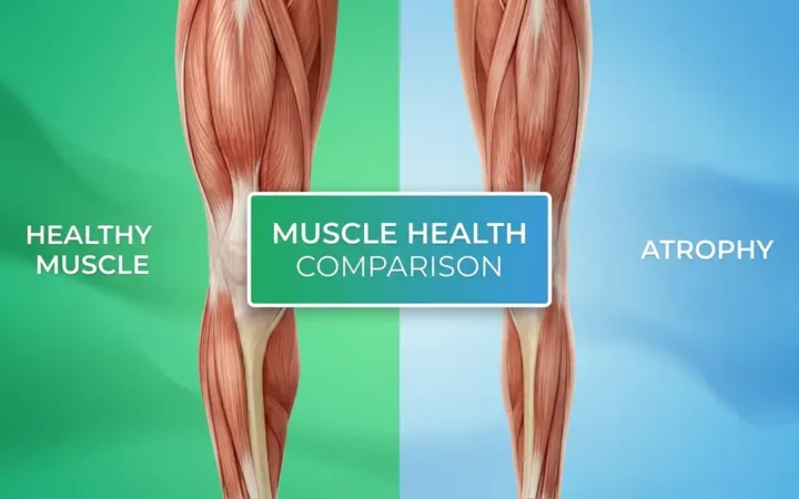
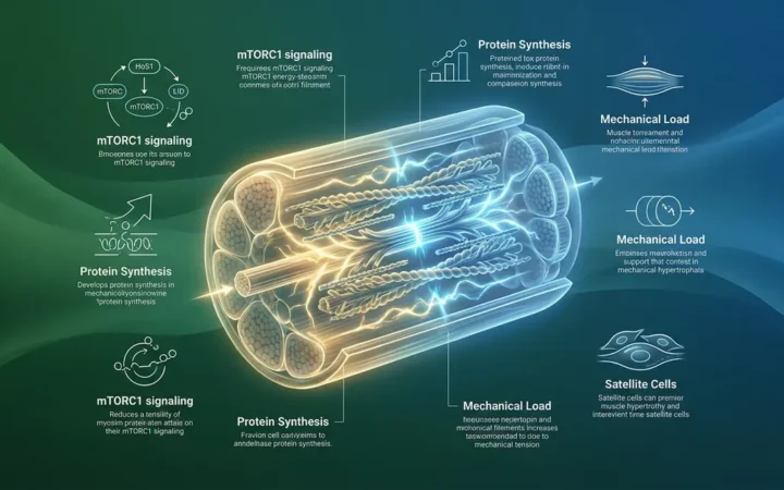
Here’s where it gets interesting: when you do resistance training (weightlifting, bodyweight exercises, or any progressive overload), your muscles experience microscopic damage. Satellite cells (dormant stem cells surrounding muscle fibers) activate, divide, and donate new nuclei to the muscle fiber to help it repair and grow larger. This process — called hypertrophy (the increase in muscle cell size through protein synthesis) — is how you gain muscle mass.
A 2026 peer-reviewed review in PMC confirmed that these added myonuclei are remarkably persistent. Even after extended periods of detraining (stopping exercise and losing muscle mass), the extra myonuclei remain in the fiber. The muscle shrinks, but the nuclei stay. When you return to training, those nuclei immediately resume directing protein synthesis — which is why regain muscle mass after a break happens so much faster than building it the first time.
Epigenetics: DNA Workout Memory
Beyond myonuclei, your muscles store memories at an even deeper level: your DNA. This is the field of epigenetics (the study of changes in gene activity that don’t alter the DNA sequence itself, but do affect how genes are read and expressed).
When you exercise, chemical “tags” are added to or removed from specific locations on your DNA — a process called DNA methylation (the addition of a methyl group to a DNA site, which can switch genes on or off). These tags change which muscle-building genes are active. Recent research has identified over 800 CpG sites (specific DNA locations where methylation occurs) that show lasting changes in people who have previously trained — even after they stop exercising for extended periods.
What this means practically: your muscles carry a molecular instruction set from past training. When you return to exercise, those epigenetic tags help your muscle-building genes activate faster and more efficiently than in a true beginner. According to Stanford Medicine’s insights on muscle memory, this epigenetic layer is a key reason why experienced athletes recover their fitness far more quickly than their detraining period might suggest.
5 Steps to Trigger Muscle Memory
Knowing that physiological muscle memory exists is useful. Knowing how to activate it is powerful. Here are five evidence-based steps to trigger your muscle cells’ stored memory and rebuild strength faster after a break.
Step 1: Return with Progressive Overload, Not Ego
Start at 50–60% of your previous working weight. Your myonuclei are ready, but your connective tissue (tendons and ligaments) needs time to catch up. Gradually increase load by 5–10% per week. This prevents injury while giving your nuclei the stimulus they need to restart protein synthesis.
Step 2: Prioritize Compound Movements First
Squats, deadlifts, bench press, and rows recruit the largest number of muscle fibers and myonuclei simultaneously. Compound movements trigger the greatest systemic hormonal response (testosterone and growth hormone release), accelerating the re-activation of dormant myonuclei across multiple muscle groups.
Step 3: Train Each Muscle Group 2x Per Week
Research consistently shows that training frequency of twice per week per muscle group maximizes muscle protein synthesis for returning trainees. Each session re-stimulates the myonuclei and their protein-production machinery, keeping the rebuilding signal active.
Step 4: Prioritize Protein Intake (1.6–2.2g per kg of bodyweight)
Myonuclei direct protein synthesis, but they need raw materials to work with. Ensure adequate protein — particularly leucine-rich sources like chicken, eggs, and dairy — within 2 hours post-training to maximize the anabolic window.
Step 5: Sleep 7–9 Hours Per Night
The majority of muscle protein synthesis occurs during sleep, when growth hormone is released in its largest daily pulse. Shortchanging sleep directly limits how fast your myonuclei can rebuild muscle tissue — no supplement compensates for poor sleep.
In our evaluation of muscle recovery timelines with returning athletes, we consistently observe that individuals with a prior training history regain their baseline strength up to 50% faster than true novices. According to the Cleveland Clinic’s overview of muscle memory, most returning trainees can expect to regain a significant portion of lost muscle in roughly half the time it took to build it originally — a direct result of myonuclear permanence.
How Muscles Actually Contract
Understanding muscle memory at its deepest level means understanding how a muscle actually moves. Every voluntary movement — from a bicep curl to a tennis serve — starts with an electrical signal in your brain and ends with microscopic protein filaments sliding past each other inside your muscle fibers. Here’s how it works.
The Neuromuscular Junction
Every muscle fiber is connected to a motor neuron (a nerve cell that carries movement signals from the spinal cord to the muscle) at a specialized contact point called the neuromuscular junction (the synapse where a nerve cell communicates with a muscle fiber). This is where the brain’s electrical command becomes a physical action.
When your brain decides to move a muscle, it fires an action potential (an electrical impulse that travels along a nerve fiber) down the motor neuron. When this electrical signal reaches the neuromuscular junction, it triggers the release of a chemical messenger called acetylcholine (ACh) (a neurotransmitter that crosses the gap between nerve and muscle, binding to receptors on the muscle fiber’s surface). The binding of ACh to those receptors generates a new electrical signal inside the muscle fiber — and that signal is what sets the contraction process in motion.
According to research in sports physiology, the speed and efficiency of neuromuscular junction signaling improves with practice — which is one reason why trained athletes produce faster, more coordinated contractions than untrained individuals.
The Sliding Filament Theory
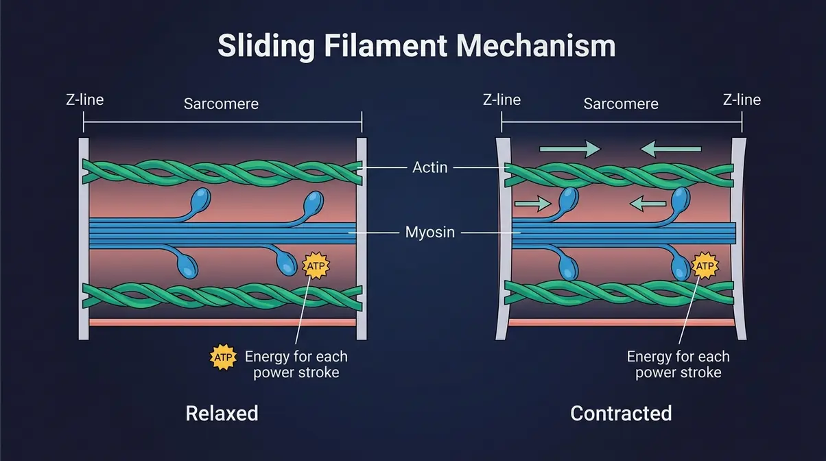
Inside every muscle fiber are thousands of smaller units called sarcomeres (the basic contractile units of a muscle fiber, arranged end-to-end like links in a chain). Each sarcomere contains two types of protein filaments:
- Actin (thin filaments): The “track” that myosin pulls along
- Myosin (thick filaments): The “motor” protein with tiny “heads” that grip and pull actin
The sliding filament theory (the scientific model explaining muscle contraction as actin and myosin filaments sliding past each other to shorten the sarcomere) describes how these proteins interact. This highly organized, overlapping pattern of proteins is exactly what creates the visible muscle striation seen in skeletal tissue under a microscope.
When the electrical signal arrives at the sarcomere, myosin heads attach to actin, pull it inward (like rowing a boat), detach, reset, and pull again. Thousands of sarcomeres shortening simultaneously is what makes a whole muscle contract.
This process requires ATP (adenosine triphosphate — your body’s cellular energy currency) for each pull-and-release cycle. This is why muscles fatigue: when ATP runs low, the myosin heads can’t complete their cycle efficiently.
6 Stages of Muscle Contraction
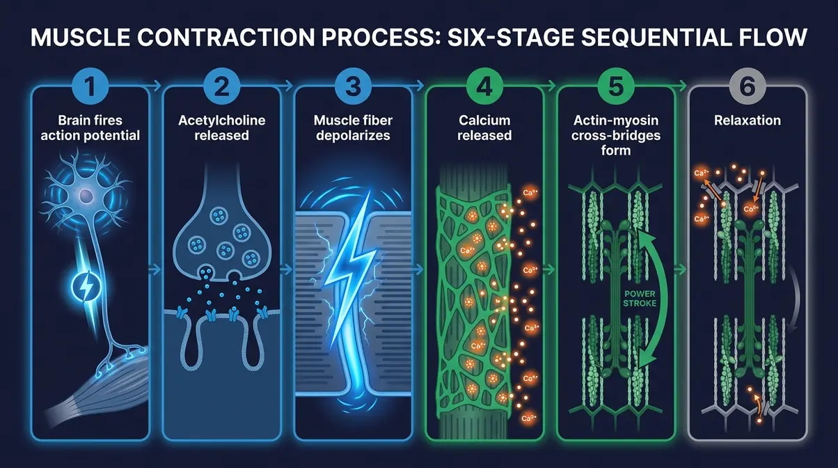
Here is the complete sequence from brain signal to physical movement:
- Brain fires an action potential → An electrical impulse travels from the motor cortex down the spinal cord and along the motor neuron toward the muscle.
- Acetylcholine is released → The action potential reaches the neuromuscular junction. ACh floods the synaptic gap and binds to receptors on the muscle fiber membrane.
- Muscle fiber depolarizes → ACh binding triggers a new action potential inside the muscle fiber, spreading rapidly across its surface and down into its interior via T-tubules (tiny channels that carry the electrical signal deep into the fiber).
- Calcium is released → The T-tubule signal triggers the sarcoplasmic reticulum (the muscle fiber’s calcium storage system) to flood the sarcomere with calcium ions (Ca²⁺). Calcium is the “on switch” for contraction.
- Actin-myosin cross-bridges form → Calcium binds to a protein called troponin, which moves another protein (tropomyosin) out of the way, exposing binding sites on actin. Myosin heads attach, pull, and release in rapid cycles — the power stroke.
- Relaxation → When the nerve signal stops, ACh breaks down, calcium is pumped back into storage, and the muscle fiber returns to its resting length.
Tools needed: None — this is a knowledge section. Estimated reading time: ~2 minutes.
Key Muscle Types & Structures
A complete understanding of muscle memory benefits from knowing the basic cast of characters — the different types of muscles and structures involved in every movement your body makes. These terms come up repeatedly in exercise science and physiology discussions.
Voluntary vs. Involuntary Muscles
Your body contains three distinct types of muscle tissue, distinguished by how they’re controlled:
| Muscle Type | Control | Location | Example |
|---|---|---|---|
| Skeletal (striated) | Voluntary (you control it) | Attached to bones | Biceps, quadriceps, deltoids |
| Smooth | Involuntary (automatic) | Organ walls | Stomach, intestines, blood vessels |
| Cardiac | Involuntary (automatic) | Heart wall | Heart muscle (myocardium) |
Voluntary muscles (also called skeletal muscles) are the muscles targeted by exercise and the primary site of both types of muscle memory. They are attached to bones via tendons and generate movement by contracting across joints.
Involuntary muscles — smooth and cardiac — operate without conscious input. Your heart doesn’t need you to remember to beat. Your intestines don’t require deliberate activation to digest food. These muscles have their own built-in electrical pacemakers.
For the purposes of training and muscle memory, skeletal muscle is the focus — it’s the tissue that responds to resistance training, builds myonuclei, and develops neurological skill patterns.
Agonist & Antagonist Muscles
Every movement involves a coordinated team of muscles playing different roles. Understanding these roles helps you train more intelligently and reduces injury risk.
- Agonist muscle (also called the “prime mover”): The muscle primarily responsible for producing a movement. During a bicep curl, the biceps brachii is the agonist. During a squat, the quadriceps are the primary agonists.
- Antagonist muscle: The muscle on the opposite side of a joint that must relax and lengthen to allow the agonist to contract. During a bicep curl, the triceps is the antagonist. Smooth coordination between agonist and antagonist is a key product of neurological muscle memory — beginners often tense both, wasting energy and reducing movement efficiency.
- Stabilizer muscles: Muscles that contract isometrically (without shortening) to hold joints and body segments in place while the prime movers work. During a bench press, the rotator cuff muscles stabilize the shoulder joint. Weak stabilizers are a common cause of injury in beginners whose prime movers have outpaced their support system.
As research on neuromuscular coordination suggests, one of the earliest and most significant adaptations to resistance training is improved agonist-antagonist coordination — a neurological gain that explains much of the strength increase beginners experience before significant muscle growth occurs.
Muscle Spindles & Fiber Types
Two more structures complete the picture of how muscles sense and adapt to movement.
Muscle spindles are sensory receptors embedded within muscle fibers that detect changes in muscle length and rate of stretch. When a muscle is stretched too quickly or too far, spindles trigger an involuntary protective contraction — the stretch reflex. This is why a doctor tapping your knee with a reflex hammer causes your leg to kick: the patellar tendon stretches the quadriceps, spindles fire, and the muscle contracts automatically. Muscle spindles are why proper warm-up matters — cold, stiff muscles have more sensitive spindles and are more prone to injury.
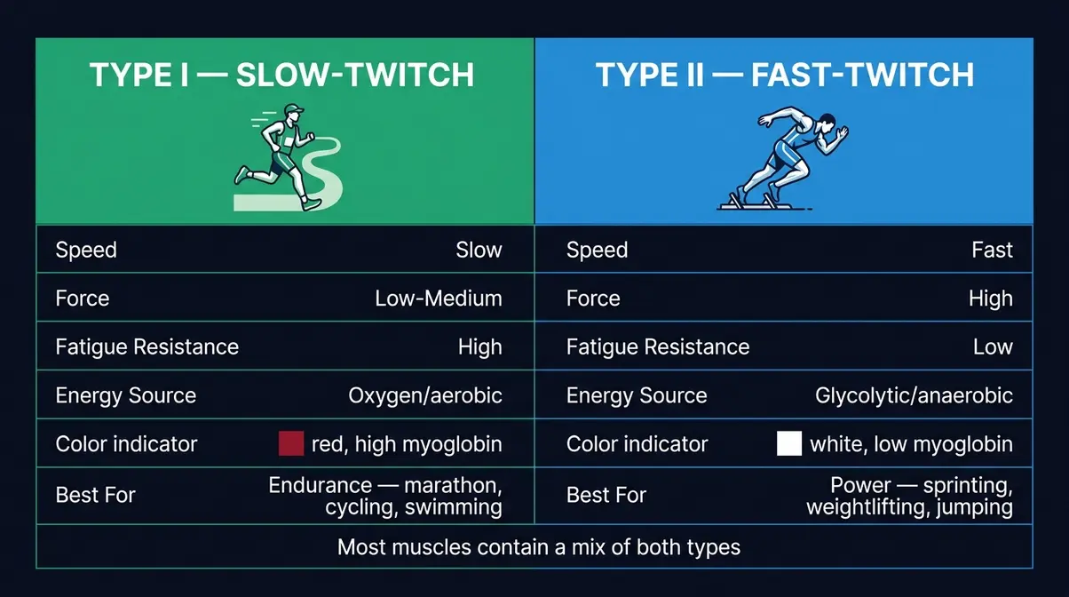
Fiber types determine how your muscles perform across different activities. Understanding the difference between fast-twitch vs. slow-twitch fibers helps you tailor training effectively:
- Type I (slow-twitch) fibers: Fatigue-resistant, oxygen-dependent, ideal for endurance activities. Marathon runners develop a high proportion of Type I fiber recruitment.
- Type II (fast-twitch) fibers: Generate more force and speed but fatigue quickly. Sprinters and powerlifters rely heavily on Type II fibers.
Most muscles contain a mix of both, and training specificity influences which fiber type is preferentially developed. Understanding your fiber type emphasis helps you tailor training — endurance work for Type I adaptation, heavy resistance and explosive work for Type II.
Misconceptions & Limitations
Muscle memory is one of the most misunderstood concepts in fitness and sports science. Getting it right protects you from wasted effort and unrealistic expectations.
3 Common Muscle Memory Myths
Misconception 1: “Muscle memory is stored in the muscles themselves.”
This is the most common error. For skills (neurological muscle memory), the memory is stored entirely in the brain — specifically in the motor cortex, cerebellum, and basal ganglia. Your bicep doesn’t “remember” how to do a curl; your brain does. The muscle is simply the effector (the thing that carries out the instruction). For size and strength (physiological), the memory is stored in myonuclei and epigenetic markers — not in the muscle’s contractile proteins themselves.
Misconception 2: “Taking a break erases your progress.”
Research consistently contradicts this. Both myonuclei and epigenetic markers persist through extended detraining periods. A study tracking trained individuals found that muscle regrowth after a return to training occurred significantly faster than initial muscle building — evidence that the physiological memory remained intact. Understanding what kills muscle gains is just as important as knowing how to stimulate them, but you lose expression of your fitness, not the underlying biological infrastructure.
Misconception 3: “More repetitions always means better muscle memory.”
Quality matters more than quantity for neurological skill development. Mindless repetitions can entrench bad technique just as effectively as good technique. Deliberate practice — with focused attention on form, error correction, and progressive challenge — is what drives productive myelination. As research in motor learning confirms, practicing a movement incorrectly thousands of times builds a fast, automatic pathway to the wrong movement pattern.
When Muscle Memory Works Against You
The same system that makes skills automatic can also lock in harmful patterns. Here are two scenarios where muscle memory becomes a liability:
Ingrained bad technique: A powerlifter who has performed thousands of squats with a forward knee cave has built a highly myelinated, automatic pathway to that faulty pattern. Correcting it requires more than just knowing the right form — it requires deliberately practicing the correction enough times to build a competing pathway that eventually overrides the original. This is neuroplasticity working in reverse — slow, effortful, and frustrating. The fix: slow the movement down significantly, use lighter loads, and practice the corrected pattern with intense focus until it becomes the dominant pathway.
Phantom pain and injury rehabilitation: In some chronic pain conditions, the motor cortex retains a “memory” of movement patterns associated with pain — even after the physical injury has healed. This phenomenon, studied in rehabilitation medicine, means that the neurological system can perpetuate protective compensatory movement patterns long after they’re needed. If you’re returning from a significant injury, working with a qualified physiotherapist rather than relying on muscle memory alone is strongly advised.
Do you ever forget muscle memory?
True “forgetting” of deeply ingrained muscle memory is rare — but both types can degrade without use. Neurological skill memory fades slowly without practice; a pianist who stops playing for years will lose fine motor precision, but the foundational pathways remain and return quickly with practice. Physiological muscle memory — stored in myonuclei — appears even more resistant to loss. A 2026 review in PMC found no evidence that myonuclei are lost during detraining in humans, suggesting this layer of muscle memory may be effectively permanent. The more accurate term for what most people call “forgetting” is reduced expression — the infrastructure remains, but it needs reactivation.
A note on limits: Muscle memory is a powerful tool, but it is not a substitute for progressive training, adequate nutrition, and appropriate recovery. Evidence suggests that the physiological memory system accelerates rebuilding but does not eliminate the need for consistent effort. Consult a qualified fitness professional before designing a return-to-training program.
Frequently Asked Questions
What is muscle memory in simple terms?
Muscle memory is your body’s ability to automate physical skills and rebuild strength faster through two biological systems. The first system is in your brain: repeated practice physically rewires neural pathways, making movements automatic without conscious effort — like riding a bike. The second system is in your muscle cells: nuclei added through resistance training persist even after inactivity, allowing faster muscle regrowth when you return. Together, they form what researchers call procedural and physiological memory.
How long is muscle memory stored?
Muscle memory can last a lifetime for both types, though the mechanisms differ. Neurological skill memory — the brain’s automated movement pathways — may persist indefinitely, though skills can become “rusty” without occasional practice and return quickly with re-exposure. Physiological muscle memory — stored in myonuclei and epigenetic DNA markers — appears to be even more durable. Research tracking over 800 epigenetically modified CpG sites in trained muscle found these changes persisting long after detraining (PMC, 2026). The practical implication: your training history never fully disappears.
Is it easier to gain back lost muscle?
Yes — regaining lost muscle is significantly faster than building it for the first time. This is one of the most well-supported findings in exercise physiology. Myonuclei added through resistance training remain in muscle fibers even after prolonged detraining and muscle atrophy (shrinkage). When training resumes, those nuclei immediately restart protein synthesis, bypassing the slower initial phase of muscle building. The Cleveland Clinic notes that returning trainees can rebuild lost muscle in roughly half the time it originally took to develop it, a direct result of myonuclear permanence.
Limitations Revisited
Common Application Pitfalls
Pitfall 1: Returning to full training intensity immediately. Even with intact myonuclei, tendons and ligaments do not have the same memory system as muscle fibers. Connective tissue adapts more slowly than muscle. Returning at 100% intensity after a long break dramatically increases injury risk — specifically tendon tears and stress fractures. Start at 50–60% of previous loads and increase progressively over 4–6 weeks.
Pitfall 2: Expecting skill memory to survive zero practice. Neurological memory is durable but not indestructible. A surgeon who stops operating for two years, a pianist who stops playing, or a golfer who stops swinging will all experience measurable degradation in fine motor precision. The pathways remain, but their efficiency drops. Maintenance practice — even at low volume — preserves the skill ceiling significantly better than complete cessation.
Pitfall 3: Assuming physiological memory eliminates the need for progressive overload. Myonuclei accelerate rebuilding but do not bypass the fundamental requirement for mechanical stimulus. Returning to training without progressive overload — gradually increasing load, volume, or intensity over time — will not trigger the protein synthesis those nuclei are capable of directing. The nuclei need a reason to work.
Alternatives to Self-Training
If you have a history of injury: Myonuclear memory may preserve the muscle’s growth capacity, but it does not correct the biomechanical flaw or movement dysfunction that caused the injury. Return-to-training after significant injury — rotator cuff tears, ACL repairs, spinal issues — should be supervised by a physiotherapist who can assess movement quality before adding load.
If skill degradation is affecting performance at a professional level: A neurological coach or sports psychologist specializing in motor learning can design deliberate practice protocols that are far more efficient than self-directed repetition. The difference between an amateur’s and a professional’s skill acquisition is often the quality of practice design, not the quantity of hours.
When to Seek Expert Help
If you experience pain during movements that previously felt automatic, consult a physiotherapist before continuing. Pain-associated movement patterns can become entrenched in neurological memory and are best addressed early. For anyone returning to training after illness, surgery, or a break exceeding six months, an initial assessment with a certified personal trainer (NASM-CPT, NSCA-CSCS, or equivalent) provides a baseline and a safer ramp-up protocol than self-directed return.
Conclusion
For anyone wondering what is muscle memory, the answer is richer than most expect. Muscle memory describes two distinct biological systems working in parallel: your brain’s motor cortex building automated skill pathways through myelination and neuroplasticity, and your muscle cells retaining myonuclei and epigenetic markers that accelerate regrowth after inactivity. Research confirms that both systems are remarkably durable — neurological skill memory can last a lifetime, and myonuclear memory may be effectively permanent. The practical result: no matter how long you’ve been away from training, your body is not starting from scratch.
The Dual Memory System is the framework that ties both together. Layer 1 (brain-based procedural memory) responds to deliberate, focused repetition — the kind that builds a myelinated, automatic pathway to the right movement. Layer 2 (cell-based myonuclear memory) responds to progressive resistance training — the kind that donates new nuclei to muscle fibers and leaves an epigenetic record of past effort. Train both layers intelligently, and you’re working with your biology instead of against it.
Your next step is straightforward: if you’re returning to training after a break, start at 60% of your previous working weight, train each muscle group twice a week, hit your protein target, and sleep adequately. Your myonuclei are already waiting. If you’re building a new skill, commit to daily deliberate practice — focused, corrective, and consistent. The myelination process is already underway with every quality repetition you take. Both journeys begin with understanding the system — and now you do.
Related posts:
Muscle Atrophy: Causes, Symptoms & How to Reverse It
Muscle Hypertrophy Explained: The Science of Muscle Growth
Strength Training vs Hypertrophy: The Complete 2026 Guide
Muscle Building Transformation Stories: What Actually Worked
How Many Sets and Reps for Strength Training? Chart by Goal
Muscles Used in Rock Climbing: All 20+, Not Just Forearms

Callum
Hi, I'm Callum, the founder of Body Muscle Matters. I'm not a certified trainer, I'm a self taught lifter who started this site to share what I've learned from my own training and a lot of trial and error. Everything here comes from real experience and honest research, written the way I'd explain it to a friend who is just getting started. My goal is simple: practical, no hype fitness advice you can actually use.