
What Is Muscle Striation? The Science and the Shredded Look
If you’ve ever looked at a bodybuilder’s chest or legs and seen what looks like steel cables running under the skin, you’ve seen muscle striations in action. But here’s what most people don’t know — those same striations exist inside every one of your muscles right now, invisible at the microscopic level. In our comprehensive review of physiological literature and NIH data, we found that understanding exactly what is muscle striation involves looking at the cellular level first.
The term sounds like advanced biology, but the confusion it causes — between the anatomy definition and the fitness goal — leaves most people with half the picture and no clear path forward. You either find a dense academic paper that skips the fitness application entirely, or a fitness article that skips the biology. Neither gives you the full story.
This guide covers the science first, then the application. You’ll learn exactly what the term means at the cellular level, how the three types of muscle tissue differ, and — if you’re chasing the super shredded, etched delts look — the evidence-based protocols for making your striations visible safely.
⚠️ Medical Disclaimer: The information in this article is for educational purposes only and does not constitute medical or fitness advice. Achieving very low body fat percentages carries real health risks. Always consult a qualified healthcare provider or certified fitness professional before making significant changes to your diet or exercise routine.
What is muscle striation? It refers to the alternating light and dark bands visible in skeletal and cardiac muscle tissue — structures present in every person’s body that power every voluntary movement you make.
- Everyone has muscle striations: They are a biological feature of skeletal and cardiac muscle, not a rare achievement reserved for bodybuilders.
- Visible striations require: Very low body fat (3–9% for men, 10–12% for women) combined with significant muscle mass built through resistance training.
- The Striation Spectrum: Striations operate on two levels — microscopic (always present in every person) and visible (requires extreme leanness to appear through the skin).
- The mechanism: Striations form from the precise, repeating arrangement of actin and myosin proteins inside structures called sarcomeres.
- Safety first: Maintaining extreme leanness long-term carries real health risks — always consult a certified professional before pursuing visible striations.
What Is Muscle Striation? Microscopic Origins
Muscle striation refers to the alternating light and dark bands visible in skeletal and cardiac muscle tissue under a microscope. These bands form because of the highly organized, repeating arrangement of two proteins — actin and myosin — inside structures called sarcomeres. Understanding what causes these stripes explains both how your muscles contract and what it takes to make them visible through your skin.
Muscle striation is not a fitness achievement — it is a fundamental structural feature of every person’s skeletal and cardiac muscle tissue, created by the precise arrangement of actin and myosin proteins inside sarcomeres. This distinction matters enormously. When most people hear the word “striations,” they picture a stage-ready bodybuilder. In reality, you already have fully formed striations inside every skeletal muscle you own — the question is only whether they’re visible.
What does muscle striation mean?
Muscle striation refers to the alternating light and dark bands visible in skeletal and cardiac muscle tissue. These bands form because of the regular, repeating arrangement of two proteins — actin and myosin — inside structures called sarcomeres, the functional units of muscle fibers. The term applies both to the microscopic anatomy visible under a microscope (present in every person) and to the visible appearance through the skin (requiring extreme leanness). In everyday fitness language, “striations” typically refers to the visible type seen in highly conditioned athletes.
The Sarcomere: Repeating Muscle Units
The sarcomere (the repeating functional unit of a muscle fiber) is the fundamental reason muscles appear striped. Think of a sarcomere like a single bead in a bracelet. Each bead looks identical, and when you line thousands of them up end-to-end, you see a repeating pattern of colors — that’s the striped muscle. Stack enough sarcomeres in a row and you form a myofibril (a single contractile strand running the length of a muscle fiber). Bundle thousands of myofibrils together and you get one muscle fiber. Your bicep alone contains millions of sarcomeres.
Inside each sarcomere, two types of protein filaments interlock in a precise arrangement. Actin (a thin protein filament) and myosin (a thick protein filament) overlap in a highly organized pattern. According to NIH review on the sliding filament history, in striated muscles, sarcomeres contain overlapping filaments that maintain their length but slide past one another during contraction (NIH, 2017). It is this overlapping, alternating arrangement that creates the banded appearance — the biological origin of striation.
Why does this matter to you? Because every time you lift a weight, sprint, or climb stairs, millions of sarcomeres in your muscles are shortening simultaneously. The striped structure you see in a bodybuilder is the same structural machinery powering your every movement — just revealed through the skin by extreme leanness.
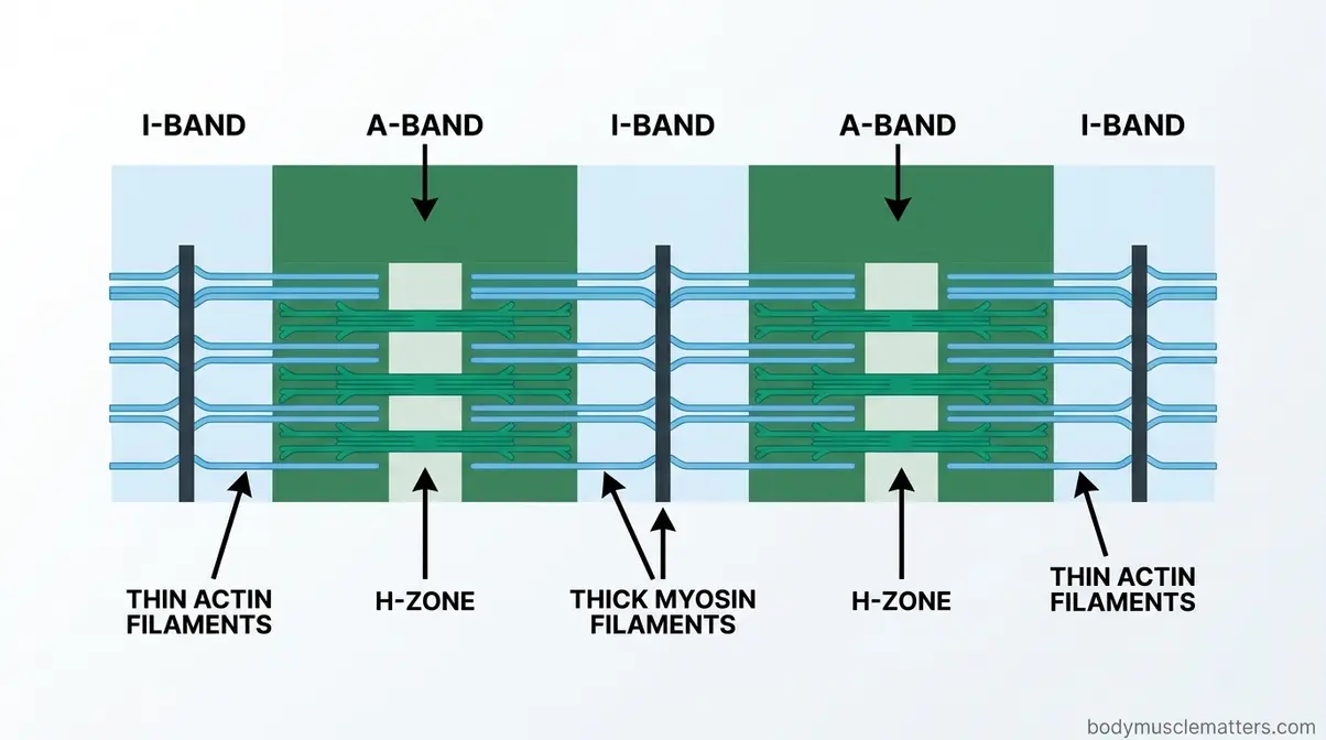
As shown in the diagram above, the alternating dark and light zones of a sarcomere have specific names that anatomy and physiology courses rely on. Understanding them gives you a precise mental model of the muscle blueprint.
A-Bands, I-Bands, and Z-Lines
The banded pattern of a sarcomere breaks down into three clearly defined structures. A simple table makes them easy to remember:
| Structure | Appearance | Contains | Function |
|---|---|---|---|
| A-band | Dark stripe | Actin + myosin overlapping | Zone of filament overlap and cross-bridge formation |
| I-band | Light stripe | Actin only (no myosin) | Region of thin filaments between sarcomeres |
| Z-line | Dense boundary line | Anchors actin filaments | Marks the border between two adjacent sarcomeres |
A memory trick that works: A = dArk (more protein mass, absorbs more light), I = lIght (fewer filaments, appears pale), Z = Zone boundary (the wall between sarcomeres). The A-band appears dark because both thick myosin and thin actin filaments overlap there, creating dense protein mass. The I-band appears lighter because only thin actin filaments are present, with less protein to absorb microscope light.
The Z-line — also called the Z-disc — acts as an anchor point for actin filaments and defines where one sarcomere ends and the next begins. Understanding these zones helps you see why the striped pattern is so regular and precise: it’s not random. It’s the same repeating structure, replicated millions of times across every muscle fiber.
This is the essence of The Striation Spectrum — the concept that muscle striations exist on two distinct levels. At the microscopic level, striations are a biological constant: every person on earth with skeletal or cardiac muscle has them, right now, regardless of body composition. At the visible level, striations only appear through the skin when body fat drops low enough for the skin to conform tightly around the muscle. You are not trying to create striations — you already have them. The only question is whether yours are visible.
Now that you can picture the microscopic stripes inside a muscle fiber, the next step is understanding which of your body’s three muscle types actually have this structure — because not all muscles are striated.
Three Muscle Tissue Types: Striated vs. Smooth
Your body contains three distinct types of muscle tissue, and only two of them are striated. According to MedlinePlus, the three types of muscle tissue are cardiac, smooth, and skeletal — each with a different structure, location, and level of conscious control (MedlinePlus, 2026). Knowing which type is which gives you a clearer picture of why striations matter in some muscles but not others.
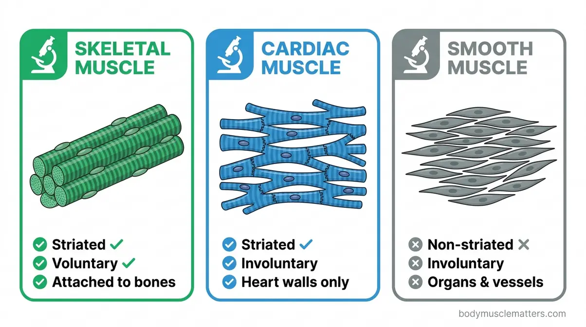
Skeletal Muscle: Voluntary Control
Skeletal muscle is the striated tissue attached to your bones that you consciously move. It is the most abundant muscle type in the body, making up roughly 40% of total body weight in a healthy adult, and it is the only type under direct voluntary control (Cleveland Clinic, 2026). Every time you curl a dumbbell, take a step, or blink, you are activating skeletal muscle.
Under a microscope, skeletal muscle fibers appear distinctly striped — a direct result of the sarcomere structure described above. Each fiber is a single, elongated cell with multiple nuclei (a feature called being multinucleated). The fibers are organized into bundles wrapped in connective tissue layers: the endomysium surrounds each individual fiber, the perimysium wraps groups of fibers into bundles called fascicles, and the epimysium encases the entire muscle. These connective tissue layers are structural details most fitness articles overlook entirely, yet they are central to how muscles transmit force to tendons and bone.
Skeletal muscle contains two primary fiber subtypes: Type I fibers (slow-twitch, highly fatigue-resistant, aerobic metabolism) and Type II fibers (fast-twitch, generate more force, fatigue more quickly). According to StatPearls via NCBI, Type I fibers are the smallest fiber type and have a high oxidative capacity suited for endurance, while Type II fibers generate rapid, powerful contractions suited for strength and speed (NCBI, 2026). Both types are striated. Both contain sarcomeres. Both contribute to the visible striations bodybuilders display on stage.
Cardiac Muscle: Involuntary Action
Cardiac muscle is striated like skeletal muscle, but it operates entirely without your conscious input. Found exclusively in the walls of the heart, cardiac muscle contracts rhythmically and continuously from before birth until death — typically 60–100 times per minute at rest, accumulating over 2.5 billion beats in an average lifetime.
Cardiac muscle cells (called cardiomyocytes) differ structurally from skeletal muscle fibers in two important ways. First, each cardiomyocyte has only one or two nuclei, not the many nuclei found in skeletal fibers. Second, cardiac cells are physically connected to neighboring cells through specialized junctions called intercalated discs, which allow electrical signals to pass rapidly from cell to cell. This is what allows the heart to contract as one coordinated unit rather than as a collection of independent fibers.
Because cardiac muscle contains sarcomeres arranged in the same actin-myosin pattern as skeletal muscle, it is also striated — you can see the characteristic bands under a microscope. However, cardiac striations are never “visible” through the skin in the fitness sense. The heart sits inside the chest wall, and no amount of body fat reduction will reveal its structure externally.
Smooth Muscle: Non-Striated Tissue
Smooth muscle is the non-striated type — and its appearance under a microscope reflects this perfectly. Where skeletal and cardiac muscle show clear alternating bands, smooth muscle cells appear uniform and spindle-shaped with no visible stripes. This is because smooth muscle lacks the organized sarcomere arrangement; its actin and myosin filaments are present but arranged in a non-repeating, irregular pattern.
Smooth muscle lines the walls of hollow organs — your stomach, intestines, blood vessels, bladder, and uterus. It operates entirely involuntarily, controlled by the autonomic nervous system and hormones. You cannot consciously flex your intestinal smooth muscle. When fitness enthusiasts talk about achieving striations, they are always referring to skeletal muscle — smooth muscle is biologically incapable of producing the visible banded appearance.
| Muscle Type | Striated? | Control | Location |
|---|---|---|---|
| Skeletal | ✅ Yes | Voluntary | Attached to bones |
| Cardiac | ✅ Yes | Involuntary | Heart walls |
| Smooth | ❌ No | Involuntary | Organs, blood vessels |
Understanding these three types makes it clear why the fitness goal of “getting striations” specifically targets skeletal muscle: it’s the only type you can build, train, and reveal through the skin. Now that you know which muscles are striated, it helps to understand precisely how those muscles contract — because the contraction mechanism is inseparable from the striation structure itself.
Muscle Contraction & Sliding Filament Theory
The sliding filament theory explains how muscles produce force: actin and myosin filaments slide past each other within sarcomeres, shortening the muscle without either filament actually changing its own length. This theory, supported by decades of peer-reviewed research, is the foundational model of muscle physiology. According to research published in the American Journal of Physiology — Cell Physiology, sarcomere mechanics during striated muscle activation are accomplished by the relative sliding of actin filaments over myosin, driven by cross-bridge cycling (Journals of Physiology, 2026). The striped structure you see in striated muscle is not decorative — it is the precise arrangement that makes this sliding mechanism possible.
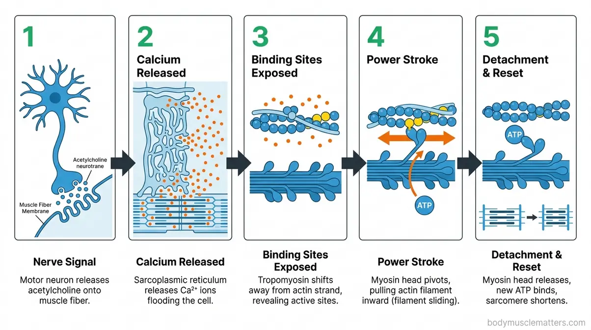
The 5-Step Contraction Process
Muscle contraction follows a precise, sequential chain of events. According to Physiology, Skeletal Muscle Contraction via NCBI Bookshelf, skeletal muscle contraction begins at the neuromuscular junction — the synapse between a motor neuron and a muscle fiber (NCBI, 2026). Here is the process broken into five clear steps:
- Nerve signal arrives. Your brain sends an electrical signal (an action potential) down a motor neuron to the neuromuscular junction. The neuron releases a chemical messenger called acetylcholine (a neurotransmitter that crosses the gap between nerve and muscle) onto the surface of the muscle fiber.
- Calcium is released. Acetylcholine triggers an electrical change in the muscle fiber’s membrane that travels into the fiber via T-tubules (tiny channels running deep into the cell). This signal causes the sarcoplasmic reticulum (the muscle’s internal calcium storage tank) to flood the cell with calcium ions (Ca²⁺).
- Binding sites are exposed. At rest, a protein called tropomyosin (a regulatory protein that blocks myosin binding sites) covers the active binding sites on actin filaments. When calcium floods in, it binds to a companion protein called troponin, which physically moves tropomyosin out of the way — exposing the binding sites on actin.
- Cross-bridges form and the power stroke occurs. Myosin heads, now able to reach actin’s exposed sites, attach and form cross-bridges. Using energy from ATP (adenosine triphosphate — the cell’s energy currency), each myosin head pivots inward, pulling the actin filament toward the center of the sarcomere. This is the “power stroke” — the moment the muscle shortens.
- Detachment and reset. A new ATP molecule binds to the myosin head, causing it to release from actin. ATP is then split, re-energizing the myosin head so it can reattach and repeat the cycle. When the nerve signal stops, calcium is pumped back into the sarcoplasmic reticulum, tropomyosin covers the binding sites again, and the muscle relaxes.
Every voluntary movement you make — from blinking to deadlifting — is this five-step sequence occurring across millions of sarcomeres simultaneously.
Actin, Myosin, and Key Molecules
The molecular players in muscle contraction each have a non-negotiable role. Understanding them helps you see why striated muscle’s structure is so specifically engineered:
- Actin (thin filament): Provides the binding sites for myosin heads. Its regular spacing within the sarcomere is what creates the light I-band.
- Myosin (thick filament): The molecular motor. Its globular heads perform the power stroke that pulls actin inward. Its dense arrangement creates the dark A-band.
- Troponin: The calcium sensor. Without it, calcium could not trigger contraction.
- Tropomyosin: The gatekeeper. It prevents unwanted contractions when the muscle should be at rest.
- ATP: The energy source. Without ATP, myosin heads cannot detach from actin — a phenomenon that explains rigor mortis (the post-death muscle stiffening that occurs when ATP production ceases and cross-bridges cannot release).
- Calcium ions (Ca²⁺): The on/off switch for the entire process.
Research from journals.physiology.org on advances in physiology education confirms that understanding these molecular roles is fundamental to grasping both normal muscle function and the basis of conditions that disrupt it (Journals of Physiology, 2026). The striped structure of muscle is not incidental — it is the precise spatial arrangement that allows these molecules to work together efficiently, step by step, contraction by contraction.
How Muscles Cooperate: Voluntary Control & Roles
Muscles rarely work in isolation. Every movement your body makes involves coordinated groups of muscles playing different roles — some contracting to produce force, others resisting or stabilizing. Understanding this coordination gives you a more complete picture of how striated skeletal muscle functions in real life, and why training programs target muscle groups rather than individual muscles.
Voluntary vs. Involuntary Muscle Control
The distinction between voluntary (consciously controlled) and involuntary (automatically controlled) muscle is one of the most practically useful in all of anatomy. Skeletal muscle operates under voluntary control — you consciously decide to reach for a cup, and your nervous system executes it. Cardiac and smooth muscle operate involuntarily, regulated by the autonomic nervous system and chemical signals without any conscious input required.
This difference traces back to the nervous system pathways involved. Voluntary skeletal muscle is controlled by the somatic nervous system, which responds to conscious commands from the motor cortex of the brain. Involuntary muscle is controlled by the autonomic nervous system, which runs continuously in the background managing heartbeat, digestion, and blood pressure. According to Cleveland Clinic, the three muscle types differ not only in structure but in the type of nerve control they receive (Cleveland Clinic, 2026). For the fitness-focused reader, this means your training targets exclusively the voluntary, striated skeletal muscle system — the one you can directly command and progressively overload.
Agonists, Antagonists, & Stabilizers
When you perform a bicep curl, your bicep is not the only muscle working. Every movement involves a coordinated team:
- Agonist (the prime mover): The muscle primarily responsible for a movement. In a bicep curl, the biceps brachii is the agonist.
- Antagonist (the opposing muscle): The muscle on the opposite side that must relax or lengthen to allow the movement. In a bicep curl, the triceps brachii acts as the antagonist.
- Stabilizers (support muscles): Muscles that don’t produce the primary movement but contract isometrically (without changing length) to hold joints steady. During a curl, your rotator cuff and core stabilize the shoulder and trunk.
- Synergists (assisting muscles): Muscles that assist the agonist. The brachialis and brachioradialis assist the biceps during elbow flexion.
Understanding agonist-antagonist pairs matters practically for training. Muscles that are chronically shortened or overworked as agonists can inhibit their antagonists — a phenomenon called reciprocal inhibition. For example, excessive sitting shortens hip flexors, which can inhibit the glutes. This is one reason why well-designed training programs deliberately balance pushing and pulling movements across all major muscle groups.
Key Muscle Structures and Terms You Should Know
Understanding a few additional anatomical terms will help you make sense of training advice, injury explanations, and physiology content you encounter as you go deeper into fitness. These terms appear constantly in workout programming, physical therapy notes, and sports science — knowing them saves you from confusion and helps you train more intelligently.
Muscle Memory, Spindles, & Fibers
Muscle memory is not the muscle literally “remembering” anything — it is a neurological phenomenon. When you repeatedly practice a movement, your nervous system becomes more efficient at recruiting the right muscle fibers in the right sequence. The motor pathways governing that movement become more myelinated (insulated) and faster. This is why a skilled weightlifter can return to training after months off and regain strength faster than a complete beginner — the neural patterns are already established.
Muscle spindles are sensory receptors embedded within skeletal muscle fibers that detect changes in muscle length and the rate of that change. When a muscle is stretched too quickly, spindles trigger the stretch reflex — a rapid, involuntary contraction designed to protect the muscle from tearing. This is the reflex that causes your knee to jerk when a doctor taps your patellar tendon. Understanding spindles explains why ballistic stretching (rapid, bouncing stretches) can trigger protective contractions that limit flexibility gains.
Regarding fiber types, recall from H2 #2 that skeletal muscle contains two primary categories. According to Physiopedia on Muscle Fibre Types, Type I (slow-twitch) fibers use aerobic respiration, produce low power output, and are highly fatigue-resistant — ideal for endurance activities like distance running (Physiopedia, 2026). Type II fibers contract faster and generate more force but fatigue quickly. Most muscles contain a mix of both types, with the ratio influenced by genetics and training history.
VMO, TVA, and Other Named Muscles
Two specific muscles come up frequently in training and rehabilitation contexts:
VMO stands for Vastus Medialis Oblique — the teardrop-shaped portion of the quadriceps visible on the inner side of the knee. It plays a critical role in knee stabilization during the final degrees of leg extension. Weakness in the VMO is commonly associated with knee pain, particularly patellofemoral pain syndrome (runner’s knee). Exercises like terminal knee extensions and narrow-stance leg presses are often prescribed to target it specifically.
TVA stands for Transversus Abdominis — the deepest layer of abdominal muscle, running horizontally around the trunk like a corset. It does not produce visible “abs” — that’s the rectus abdominis — but it creates intra-abdominal pressure that stabilizes the lumbar spine during heavy lifts. Activating the TVA (“bracing” the core) before a deadlift or squat is a fundamental technique cue in strength training because it protects the lower back.
Both the VMO and TVA are skeletal muscles — striated, voluntary, and trainable through targeted exercise.
Common Muscle Conditions and States
Muscles don’t always function optimally. Injuries, overuse, neurological issues, and recovery states all affect how striated skeletal muscle performs — and knowing the terminology helps you communicate clearly with trainers, physiotherapists, and healthcare providers. Here is a plain-English guide to the most commonly encountered muscle conditions.
Contracture, Stiffness, & Inhibition
A muscle contracture is a permanent or semi-permanent shortening of a muscle or connective tissue, resulting in reduced range of motion at a joint. Unlike a temporary cramp (which resolves quickly), a contracture persists at rest and does not release with relaxation. Contractures commonly develop after prolonged immobilization, severe burns, neurological conditions like cerebral palsy, or following untreated muscle injuries. If you are dealing with persistent tightness, learning How to Speed Up Muscle Strain Recovery can help prevent long-term contractures. According to Kenhub’s overview of muscle cell types, structural changes in muscle tissue can significantly alter its mechanical properties and function (Kenhub, 2026).
Muscle stiffness — the sensation of tightness and resistance to movement — is far more common and usually temporary. It arises from delayed onset muscle soreness (DOMS) after unfamiliar exercise, prolonged static postures, dehydration, or inadequate warm-up. DOMS peaks 24–72 hours after exercise and results from microscopic damage to muscle fibers and surrounding connective tissue, triggering an inflammatory response that the body uses to repair and strengthen the tissue. Understanding these mechanisms is also key when figuring out What Are Muscle Knots that Crunch With Massage?
Muscle inhibition (also called arthrogenic muscle inhibition) occurs when pain or injury at a joint triggers the nervous system to reduce activation of surrounding muscles as a protective mechanism. A common example: swelling in the knee after injury reflexively inhibits the quadriceps, causing noticeable weakness even when the muscle itself is undamaged. Addressing inhibition often requires specific activation exercises before strength training can resume effectively.
Activation, EMS, and Recovery States
Muscle activation refers to the process of recruiting motor units (groups of muscle fibers controlled by a single motor neuron) to produce force. In practice, “activation” exercises are low-intensity movements performed before a workout to prime specific muscles — particularly those prone to inhibition, like the glutes. A set of banded clamshells before squatting, for instance, activates the gluteus medius to improve hip stability during the main lift.
Electromuscular stimulation (EMS) — also called electrical muscle stimulation — uses low-level electrical current applied to the skin to trigger muscle contractions without voluntary neural input. EMS devices are used in physical therapy for rehabilitation of injured or inhibited muscles, and increasingly in sports performance settings to supplement training volume. Research on EMS as a standalone training tool shows modest benefits for muscle strength in clinical populations, but it does not replace progressive resistance training for hypertrophy in healthy individuals.
Recovery states matter as much as training. Muscle repair occurs primarily during rest — specifically during sleep, when growth hormone secretion peaks and protein synthesis rates are elevated. The practical takeaway: inadequate sleep directly impairs the muscle repair process, regardless of how well you train or eat. Evidence from peer-reviewed literature consistently links 7–9 hours of sleep per night with optimal recovery and performance outcomes.
Achieving Visible Muscle Striations: Protocols
Now the biology connects directly to the fitness goal. Achieving visible muscle striations requires satisfying two simultaneous prerequisites — and most people underestimate how demanding both conditions are. This section translates the sarcomere science into actionable, evidence-based guidance. Consult a certified fitness professional or healthcare provider before pursuing the extreme leanness described below.
“They’re after the ‘striated’ look, and there’s exactly one way to get it: Drop your body fat percentage to between two and five percent.”
This quote captures the reality facing competitive bodybuilders. But as you’ll see, the visible striation threshold is wider than that for most people — and the protocols to reach it are specific and measurable.
How do you get muscle striations?
Getting visible muscle striations requires two simultaneous conditions: significant muscle mass and very low body fat. Research supports a structured approach: build a hypertrophy foundation through 12–18 months of progressive resistance training, then reduce body fat through a moderate caloric deficit (300–500 calories below TDEE) with high protein intake (1.6–2.2g per kg of body weight). Cardio accelerates the deficit. The process typically takes years, not weeks, and the final body fat levels required (3–9% for men, 10–14% for women) carry real health risks — always work with a certified professional.
Prerequisites: Muscle Mass & Low Fat
The Striation Spectrum makes the prerequisites clear. Striations already exist microscopically in every skeletal muscle you have. To make them visible through the skin, you need two things working simultaneously:
Prerequisite 1: Sufficient muscle mass. Striations become visible when muscle tissue pushes outward against skin that sits close to the surface. A person at 7% body fat with very little muscle mass may show skin texture but not the distinct “feathered” or “etched” appearance. The muscle must be developed enough to create three-dimensional depth — the peaks and valleys that light catches to reveal definition. This is why visible striations are rare in untrained individuals even at low body fat: there is not enough muscle volume to create the contrast.
Prerequisite 2: Very low body fat. Fat tissue sits between the skin and the muscle. Even a thin layer of subcutaneous fat (fat directly under the skin) blurs the outline of the muscle beneath it. As body fat decreases, the skin “shrinks” closer to the muscle surface, eventually conforming tightly enough that the individual muscle heads — and ultimately the sarcomere-based banding — become visible through the skin.
Research published in PMC via NIH on body composition assessment confirms that the relationship between fat mass and muscle visibility is direct — reduced subcutaneous fat is the primary driver of increased muscle definition at any given muscle mass level (PMC, 2026). Building muscle and reducing fat are not competing goals; they are both required.
Body Fat Thresholds for Striations
Body fat thresholds for visible striations vary by individual genetics, muscle distribution, and skin thickness — but research and bodybuilding community consensus establish reliable ranges:
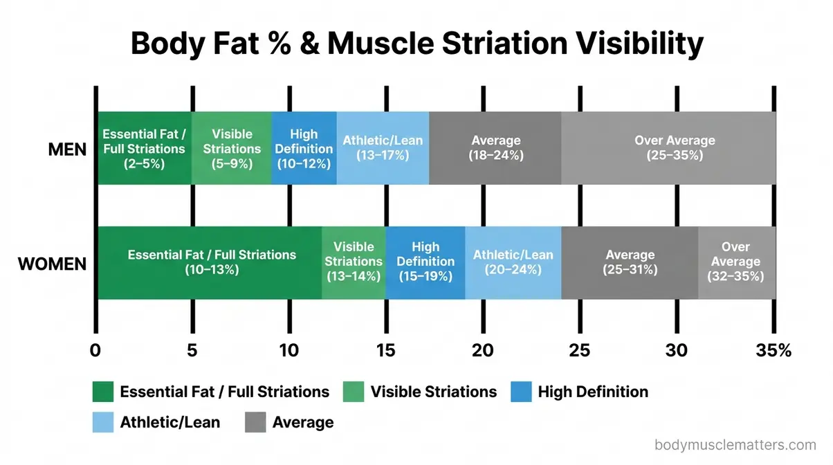
| Body Fat Range | Men | Women | Striation Visibility |
|---|---|---|---|
| Essential fat zone | 2–5% | 10–13% | Full striations across most muscle groups; seen in competitive bodybuilders |
| Visible striations | 5–9% | 10–14% | Clear striations in developed muscle groups; vascularity prominent |
| High definition | 10–12% | 15–19% | Muscle separation visible; striations appear on contraction, not at rest |
| Athletic/lean | 13–17% | 20–24% | Muscle definition visible; no resting striations |
| Average | 18–24% | 25–31% | Minimal definition; striations not visible |
According to InBody USA’s body fat percentage research, essential fat for men ranges from 2–5% of total body weight, while women require 10–13% for normal hormonal function and reproductive health (InBody USA, 2026). Dropping below essential fat thresholds — even briefly — carries significant health risks detailed in the Limitations section below.
Genetics play a meaningful role. Some individuals display visible striations in the quadriceps at 8–9% body fat, while others require 5–6% to achieve the same appearance in the same muscle group. Skin thickness, muscle belly shape, and the distribution of subcutaneous fat across the body all vary between individuals and are substantially determined by genetics.
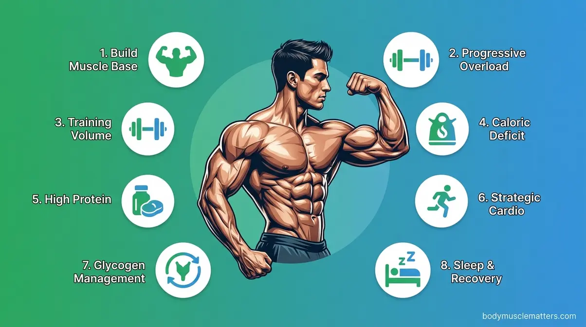
8 Protocols to Reveal Striations
These eight protocols are grounded in peer-reviewed evidence and represent the established approach used by competitive natural bodybuilders and physique athletes. Apply them progressively, and always within the guidance of a certified professional.
1. Build a Muscle Mass Foundation First
Visible striations require muscle volume. Prioritize hypertrophy (muscle growth) training before aggressive fat loss phases. For a comprehensive approach to the first prerequisite, follow a structured program like our How to Build Muscle: 9-Step Science-Backed Guide. According to research published in PMC (2026), resistance training in a caloric deficit allowed 85% of participants to gain fat-free mass simultaneously — but this works best when a foundation of muscle already exists. Aim for at least 12–18 months of consistent resistance training before pursuing extreme leanness.
2. Apply Progressive Overload
Progressive overload — gradually increasing the training stimulus over time by adding weight, reps, or sets — is the primary driver of muscle hypertrophy. According to University Hospitals on progressive overload, systematically increasing training demands keeps muscles challenged and growing across all fitness levels (University Hospitals, 2026). Track your lifts and aim for incremental improvements each week or training block.
3. Train With Sufficient Volume
Evidence-based hypertrophy guidelines recommend 10–20 working sets per muscle group per week, performed in the 6–20 repetition range, taken close to muscular failure. Compound movements — squats, deadlifts, rows, presses — provide the most efficient volume. Isolation exercises add targeted detail work for specific muscle groups like the VMO or rear deltoids.
4. Implement a Moderate Caloric Deficit
Fat loss requires a sustained caloric deficit. Research supports a moderate deficit of 300–500 calories per day below your total daily energy expenditure (TDEE) as the optimal range — aggressive enough to drive fat loss but conservative enough to preserve muscle mass. Deficits exceeding 750–1,000 calories per day accelerate muscle loss alongside fat loss, undermining the first prerequisite. To ensure you don’t lose the muscle you’ve built, read our guide on How to Prevent Muscle Loss When Cutting.
5. Prioritize Protein Intake
High protein intake preserves muscle during fat loss phases. Current evidence recommends 1.6–2.2 grams of protein per kilogram of body weight per day for individuals in a caloric deficit who are resistance training. Distributing protein intake across 4–5 meals throughout the day maximizes muscle protein synthesis compared to consuming the same total in one or two sittings.
6. Incorporate Strategic Cardio
Cardiovascular exercise accelerates the caloric deficit without further reducing food intake. Low-intensity steady-state cardio (LISS) — 30–45 minutes of walking, cycling, or rowing — is effective for fat loss with minimal impact on muscle recovery. High-intensity interval training (HIIT) provides a greater caloric burn in less time but demands more recovery. Balance cardio volume against resistance training recovery needs.
7. Manage Water and Glycogen Strategically
In the final days before a competition or photoshoot, competitive bodybuilders manipulate water and glycogen (stored carbohydrate in muscle) to maximize the appearance of striations. Proper glycogen loading causes muscles to appear fuller and more defined, while strategic water management reduces subcutaneous fluid. This is a short-term, highly specific technique — not a sustainable or health-recommended approach for everyday fitness goals.
8. Allow Sufficient Recovery and Sleep
Muscle repair — and therefore muscle growth — occurs during rest. Research consistently links 7–9 hours of sleep per night with optimized muscle protein synthesis, hormonal balance (particularly growth hormone and testosterone), and performance recovery. Chronically insufficient sleep elevates cortisol (a catabolic hormone that promotes muscle breakdown) and suppresses anabolic hormone output, directly working against the muscle-building prerequisite for visible striations.
Limitations & Safety of Extreme Leanness
The protocols above work — but the body fat levels required for visible striations carry real physiological costs. This section is not meant to discourage you from having ambitious fitness goals; it is meant to ensure you pursue them with accurate information and appropriate professional support.
Common Pitfalls and Health Risks
Pitfall 1: Hormonal disruption. The American College of Sports Medicine (ACSM) acknowledges a healthy body fat range of 10–22% for men and 20–32% for women. Dropping significantly below these ranges — particularly into the 3–6% range for men and below 13% for women — disrupts hormone production. According to InBody USA’s analysis of low body fat health effects, extremely low body fat suppresses testosterone in men and estrogen in women, impairing immune function, bone density, and reproductive health (InBody USA, 2026). The specific scenario to watch for: feeling constantly cold, losing libido, or experiencing unusual fatigue at very low body fat — these are physiological warning signals, not signs to push harder.
Pitfall 2: Female Athlete Triad / RED-S. Women face a specific cluster of risks at very low body fat, collectively called the Female Athlete Triad: low energy availability, menstrual dysfunction (amenorrhea — the cessation of periods), and reduced bone mineral density. The broader framework, now called Relative Energy Deficiency in Sport (RED-S), applies to both men and women and encompasses hormonal, psychological, and performance consequences of chronic energy deficit. Amenorrhea is not a sign of extreme fitness — it is a medical signal that the body has entered an energy crisis.
Pitfall 3: Bone density loss. Research published in Healthline citing a study of 1,767 premenopausal women found that women with low body weight showed significantly higher rates of low bone mineral density compared to those at healthy weight (Healthline, 2026). Bone density lost during periods of extreme leanness is not always fully recovered.
Pitfall 4: Psychological risk. Pursuing extreme leanness can intersect with disordered eating patterns. Rigid food tracking, fear of eating certain foods, and identity tied to body fat percentage are risk factors for orthorexia and other eating disorders. If your pursuit of visible striations is affecting your relationship with food or your quality of life, consult a mental health professional alongside a registered dietitian.
When to Choose Alternatives
Visible muscle striations are not the only marker of impressive physical development or good health. Consider these alternatives:
- If your goal is health: Target the ACSM’s recommended healthy body fat ranges (10–22% for men, 20–32% for women). Muscle definition, improved strength, and better metabolic markers are achievable at these levels without the risks of extreme leanness.
- If your goal is athletic performance: Most elite athletes perform optimally at body fat levels well above competition bodybuilding ranges. Strength, speed, and endurance do not require visible striations.
- If you have a history of disordered eating: Work with a registered dietitian and therapist before pursuing a fat loss phase. The psychological demands of extreme leanness can trigger or worsen disordered eating patterns.
When to Seek Expert Help
Consult a certified fitness professional or healthcare provider before beginning any fat loss phase targeting body fat below 10% (men) or 18% (women). Seek immediate medical attention if you experience hormonal symptoms (loss of menstrual cycle, severe libido changes), persistent fatigue, stress fractures, or significant mood disturbances during a cutting phase. These are not minor inconveniences — they are physiological signals that require professional evaluation.
Muscle Striation FAQs
What do visible striations mean?
Seeing muscle striations means your body fat is low enough for the skin to conform tightly against the underlying muscle tissue. At this point, the individual muscle heads and the three-dimensional surface of the muscle become visible through the skin. It indicates both a high degree of muscular development and very low subcutaneous fat — typically 3–9% body fat for men and 10–14% for women, according to InBody USA.
What body fat shows striations?
For most men, visible muscle striations begin appearing at approximately 7–9% body fat in well-developed muscle groups. Full striations across multiple muscle groups typically require 3–6% body fat. For women, striations begin appearing at approximately 12–14% body fat due to the higher essential fat requirement for hormonal health — women need a minimum of 10–13% body fat for normal physiological function (InBody USA, 2026). Individual variation is significant based on genetics and muscle development.
How rare are muscle striations?
Visible muscle striations through the skin are quite rare in the general population. While microscopic striations are universal, the extreme leanness required to see them is not. Visible striations require body fat levels that fewer than 1–2% of the population sustain outside of competitive bodybuilding preparation, based on general health statistics.
Are muscle striations good?
Muscle striations at the microscopic level are not just “good” — they are essential. The sarcomere structure that creates striation is the same structure that powers every voluntary movement you make. At the visible level, however, the extreme body fat levels required to sustain them carry hormonal, reproductive, and bone density risks.
What muscle is hardest to grow?
The calves (gastrocnemius and soleus) are widely considered the most difficult muscles to grow for most people. This is partly due to genetics — calf muscle insertion points and fiber type ratios are largely inherited. It is also due to their high Type I (slow-twitch, fatigue-resistant) fiber content, which requires higher training volumes to stimulate hypertrophy.
What does striation mean?
Striation means a series of parallel grooves, lines, or bands — a pattern of alternating stripes. In biology and anatomy, striation refers to the banded appearance of muscle tissue caused by the organized arrangement of protein filaments. The word comes from the Latin stria, meaning groove or furrow.
Conclusion
For anyone exploring the science of muscle — from the curious student to the goal-oriented gym-goer — muscle striation is one of the most instructive concepts in all of human biology. When asking what is muscle striation, remember it describes the alternating light and dark bands in skeletal and cardiac muscle tissue, formed by the precise repeating arrangement of actin and myosin proteins within sarcomeres. Research from NIH, the American Journal of Physiology, and Cleveland Clinic consistently confirms that this structure is universal — every person has it — and that it is the direct mechanical basis for every voluntary movement the human body produces.
The Striation Spectrum reframes the entire conversation. You are not trying to build something that doesn’t exist. Your striations are already there, at the microscopic level, powering your body right now. The question is only whether they’re visible — and the answer depends on two trainable, measurable variables: muscle mass and body fat percentage. Achieving the visible end of the Striation Spectrum takes years of consistent work, precise nutrition, and careful attention to the health risks of extreme leanness.
Start by building your muscle foundation through progressive resistance training, prioritizing compound movements and consistent progressive overload. If visible striations are your goal, work with a certified strength and conditioning specialist or registered dietitian to set realistic body fat targets and a safe, structured timeline. Consult your healthcare provider before any significant dietary change. The biology is on your side — now the plan is yours to build.
Related posts:
StrongLifts 5x5 for Beginners: Complete Guide 2026
How to Get a Bigger Bum Without Exercise: 3-Tier Guide
What Muscles Does the StairMaster Work? 6 Groups
Olympic Weightlifting vs Powerlifting: 2026 Guide
What Kills Muscle Gains? 7 Proven Saboteurs to Fix Now
Best Strength Training Apps for Women: Top 7 Picks
Callum
Hi, I'm Callum, the founder of Body Muscle Matters. I'm not a certified trainer, I'm a self taught lifter who started this site to share what I've learned from my own training and a lot of trial and error. Everything here comes from real experience and honest research, written the way I'd explain it to a friend who is just getting started. My goal is simple: practical, no hype fitness advice you can actually use.